



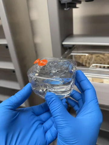
Our ophthalmology services play a pivotal role in preclinical drug development in rodents, serving to evaluate potential treatments for a wide range of eye diseases and conditions. Rodent models provide valuable insights into the mechanisms of retinal diseases, glaucoma, and other ocular disorders. Through detailed ocular examinations and imaging techniques, coupled with precise drug product delivery, we provide the framework to our sponsor partners to assess drug efficacy, ocular distribution, and possible adverse effects on the eye's structures.
Delivery Modalities
Intraorbital (IO): An orbital delivery approach for selected ocular or optic-nerve research, with the anatomical target and procedure defined for the study.
Subretinal (SR): Delivery into the subretinal space for an appropriate ocular research question.
Intravitreal (IVT): Administration into the vitreous cavity, with the procedure and test article defined for the study.
[if!supportLists]· Intracameral (IC) - Intracameral delivery involves injecting substances into the front chamber of the eye.
Behavioral Assays
Optomotor response
Assesses visual function and acuity by measuring the reflexive head and neck movements of rodents in response to rotating stripes. This test is useful for studying visual perception and neurodegenerative diseases affecting vision.
Optical coherence tomography (OCT)
Optical coherence tomography imaging is a valuable tool in preclinical drug development, for assessing retinal and ocular health. OCT provides high-resolution, cross-sectional images of the retina, allowing for detailed structural analysis. This non-invasive imaging technique is critical for evaluating the effects of pharmacological interventions on the eye and for studying the progression of ocular diseases.
In vivo electrophysiology
Electroretinography (ERG) records retinal electrical responses to light. MuriPhys uses the Celeris light-guide electrode-stimulator platform for study-appropriate flash or pattern ERG work. The recording configuration, controls and analysis are selected for the retinal functional question.